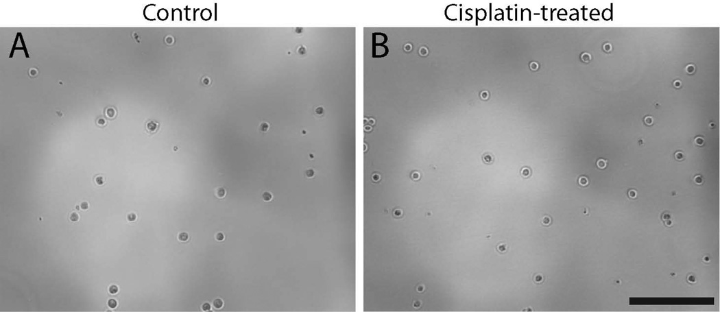
Figure 4. Normal-appearing cells can be harvested from the utricular sensory epithelium immediately following cisplatin treatment.
Utricles were placed in organotypic culture and maintained for 24 hr in either 10 µM cisplatin or in cisplatin-free medium (controls). Specimens were then rinsed and the sensory epithelia were removed via thermolysin treatment. Isolated epithelia were pooled in groups of eight, incubated in tryspin, and dissociated via gentle trituration. We observed no differences in cell appearance in untreated specimens (A) vs. the cisplatin-treated specimens (B.) In both groups, the approximate density of viable cells (as identified by trypan blue labeling) was ~3 × 10⁴ cells/ml. Scale bar = 100 µm.

