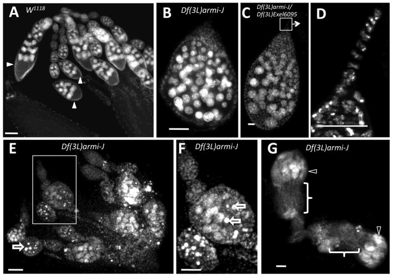
Figure 2.

Deletion of both armi and CycJ results in accumulation of egg chambers with excess germline cells. Ovaries from wild type (w1118) and Df(3L)armi-J mutant females stained with DAPI to visualize nuclei. (A) Wild-type ovarioles from 3-day old or 8-day old females (not shown) consist of chains of developing egg chambers (arrows). Each egg chamber has 15 nurse cells undergoing endoreplication (large nuclei) and a single oocyte located at the posterior end (arrowheads). (B) A typical cyst from 3-day old Df(3L)armi-J females has many more than 16 nuclei undergoing endoreplicative cycles along with pycnotic nuclei. (C) An ovariole from a Df(3L)armi-J/Df(3L)Exel6095 transheterozygote, which have a phenotype identical to Df(3L)armi-J. (D) A higher magnification of the terminal filament region in C. (E) Ovaries from 3-day old Df(3L)armi-J females contain abnormal egg chambers with more than the normal number of endoreplicating nuclei. Some cells appear to be undergoing cell death as indicated by the characteristic pycnotic nurse cell nuclei (open arrows), which is evident at higher magnification in (F). (G) Ovaries from 8-day old Df(3L)armi-J females contain empty ovarioles (bracket) with disorganized germaria (open arrowheads). Anterior is generally toward the top of each figure; in (G) the anterior of the lower ovariole is toward the right. Size bar is 50 µm in all panels except E and G, in which it is 20 µm.
