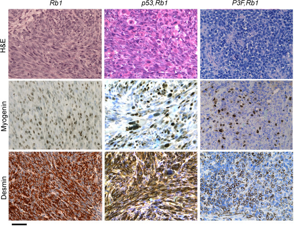
Figure 4.

Histological analysis of rare Rb1 and Pax3:Foxo1a,Rb1 tumors. A representative Rb1 tumor (left) shows spindle cell morphology with high percentage of myogenin-positive and desmin-positive cells consistent with eRMS, whereas a representative Pax3:Foxo1a,Rb1 tumor (right) consists of small round blue cells that are only rarely myogenin and desmin positive (best region shown in figure), consistent with the diagnosis of poorly differentiated malignant epithelioid neoplasm. Scale bar: 40 μm. H&E, hematoxylin and eosin.
