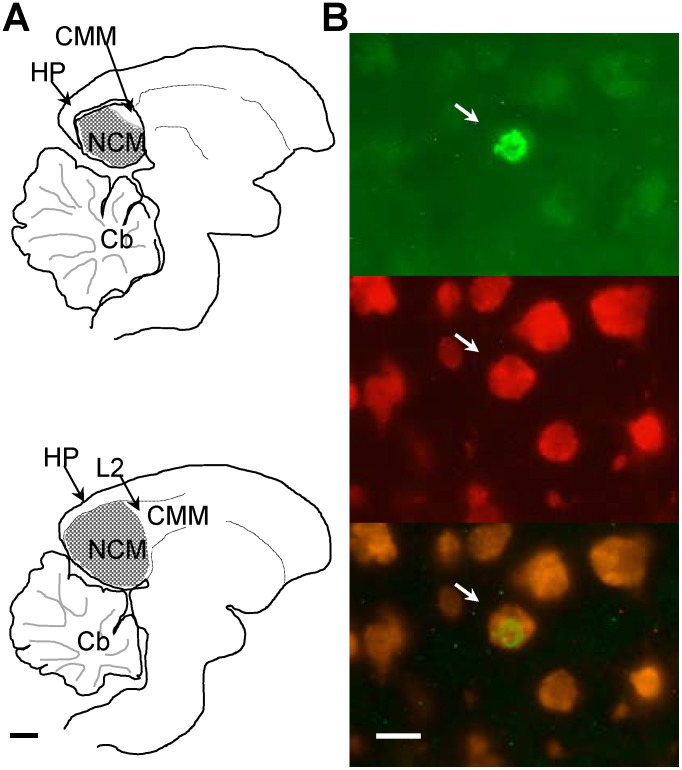
Figure 2. New neurons in NCM.
(A) Medial (top, ∼170 µm from the midline) and lateral (bottom, ∼500 µm from the midline) sections of NCM (stippled area), showing the region where new neurons were quantified. Abbreviations: Cb, cerebellum; CMM, caudomedial mesopallium; HP, hippocampus; L2, Field L2; NCM, caudomedial nidopallium. Scale bar = 1 mm. (B) Photographs of a 1-month-old new neuron in NCM (arrow) in the same field of view showing a BrdU+ nucleus (top), Hu+ neuronal cytoplasm (middle) and co-localization of both markers (bottom). Scale bar = 10 µm.

