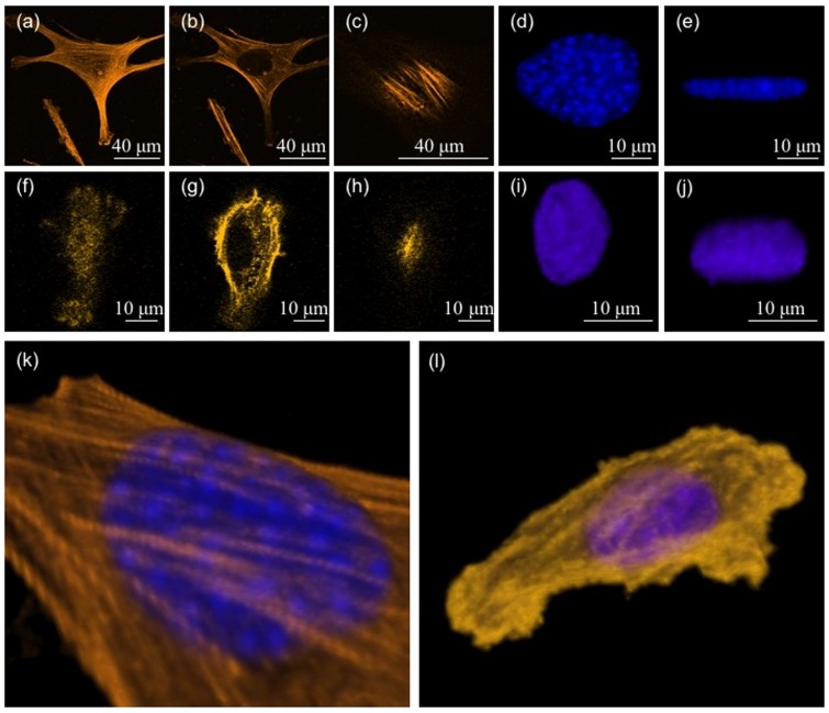
Figure 7. Confocal images showing the actin distribution and nuclear shape in cells spread on gels of two different stiffnesses.
(a, b, c) Confocal images of a cell on a 70 kPa stiffness substrate. The images show actin filaments close to the plane of the substrate, in an approximate mid plane, and just above the nucleus respectively. (d, e) The nucleus of the same cell projected in the plane of the substrate and in a perpendicular plane respectively. (f–j) Similar observations of a cell spread on a soft substrate (3 kPa). Note the difference in the nuclear shape compared to the upper set. (k, l) 3D reconstruction of confocal images showing perinuclear stress fibers running over the nucleus in the case of the first cell (stiff substrate) and a predominantly cortical actin mesh in the case of the second cell (soft substrate). Images in k and l are 3D reconstructions of the cells shown in a–e and f–j respectively.

