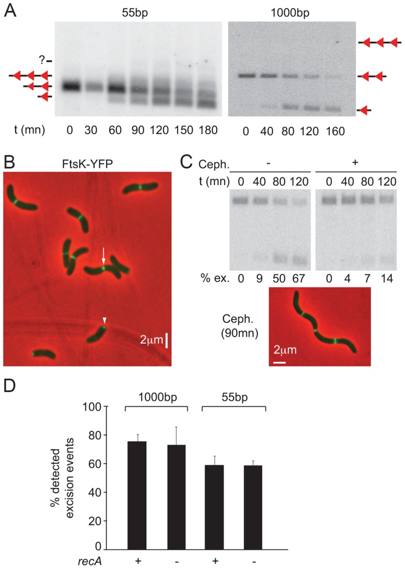
Figure 3. Control of dif-recombination.
A. Structural control of Xer recombination. Southern blot showing the different recombination products obtained with 55 bp- and 1 kbp- cassettes inserted at the dif1 locus. B. FtsK-YFP localization. The white arrow indicates a cell in which FtsK is located at the septum; the white arrowhead shows a cell in which FtsK is located at the new pole. C. Temporal control of Xer recombination. Upper panels: southern blot showing the excision of a 1 kbp cassette inserted at the dif1 locus, without or with cephalexin treatment. t: time of the experiment; ex.: excision frequency. Lower panel: snapshot showing that cephalexin treatment results in filamentation but does not prevent FtsK localization to mid-cell. D. RecA-independent recombination between dif2 sites inserted at the dif1 locus.

