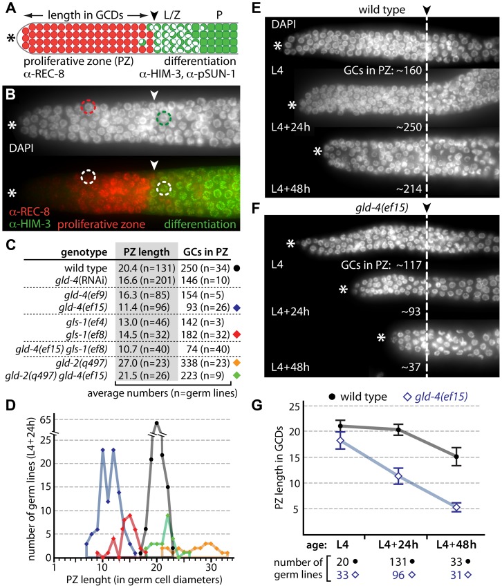
Figure 1. gld-4 and gld-2 have opposing functions in regulating the balance between proliferation and differentiation.
(A) Diagram of the distal end of an adult germ line (not to scale). Germ cells divide in the proliferative zone (PZ; red circles) and express REC-8 prior to meiotic prophase entry. Germ cells in meiotic prophase (green) express HIM-3 and pSUN-1; leptotene/zygotene (L/Z; half-filled circles) and pachytene (P; squares). In adulthood, a mitosis-to-meiosis boundary (arrowhead) is maintained at a defined distance from the distal tip (asterisk). GCDs, germ cell diameters. (B) Corresponding immunofluorescence micrograph of a distal gonad, illustrating a typical PZ nucleus (red circle) and a crescent-shaped meiotic prophase nucleus (green circle). The mitosis-to-meiosis boundary is apparent from REC-8 and HIM-3 expression. (C,D) Proliferative zone measurements of cytoPAP mutant gonads reveal germ cell (GC) number differences in young adults (L4+24 h). Circle and diamonds to the right side of the table in (C) correspond to the data shown in (D). (E,F) Age-dependent PZ changes are enhanced in gld-4 mutants. Dashed line, mitosis-to-meiosis boundary. (G) Quantification of PZ length changes of E and F over time. Error bars, standard deviations.

