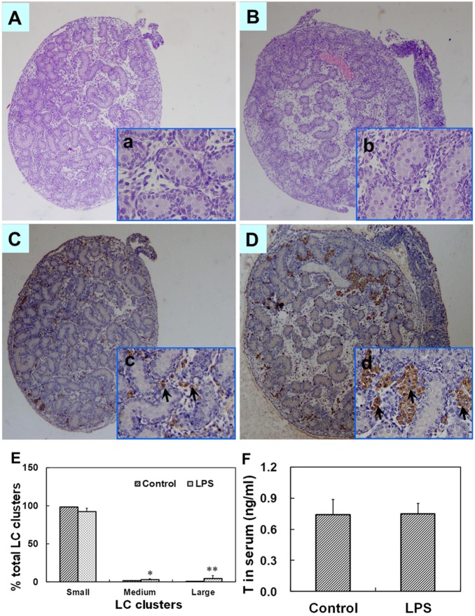
Figure 3. Testicular histology and serum T in male fetuses at GD 18.
Maternal mice were injected with LPS (50 µg/kg, i.p.) daily from gestational day (GD) 13 to GD18. The dams were sacrificed on GD 18. Testes were collected from male fetuses at 6 h after the last LPS treatment. Testicular cross-sections from controls (A and a) and LPS-treated mice (B and b) were stained with H&E. (A and B) Magnification 50×; (a and b) Magnification 200×. Leydig cells in fetal testes from controls (C and c) and LPS-treated mice (D and d) were immunolocalized by staining with a polyclonal antibody against 3β-HSD. Arrows show 3β-HSD-positive cells. (C and D) Magnification 50×; (c and d) Magnification 200×. (E) Distribution of Leydig cell (LC) clusters in fetal testes was analyzed. Small clusters accounting for ≤5% of total LC cluster area per testis, medium clusters for 5.1–14.9% and large clusters for ≥15% of total LC cluster area per testis. (F) Serum T in male fetuses was measured by RIA. Data were expressed as means ± SEM. *P<0.05, **P<0.01 as compared with controls.

