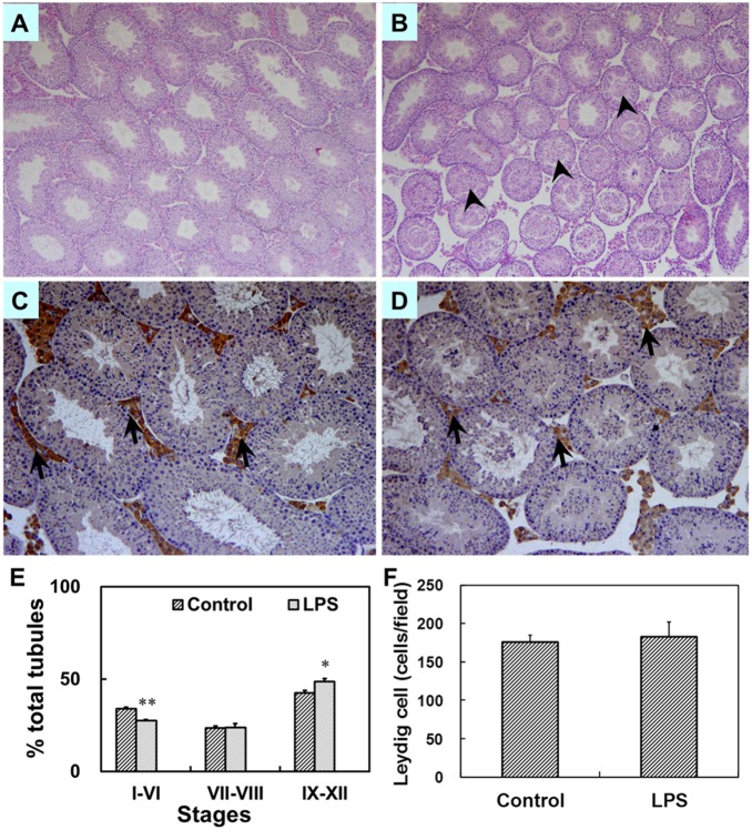
Figure 8. Testicular histology in male mice at PND 63.
Maternal mice were injected with LPS (50 µg/kg, i.p.) daily from gestational day (GD) 13 to GD18. Within 24 h after birth, excess pups were removed, so that four males and four females were kept per dam. At postnatal day (PND) 21, pups were separated from the siblings and housed five to a cage. Some males each group were sacrificed on PND 63. Testes were collected from male offsprings. Testicular cross-sections from controls (A) and LPS-treated mice (B) were stained with H&E at magnification 50×. Arrowheads show massive sloughed germ cells in the lumen of tubules. Leydig cells in testes from controls (C) and LPS-treated mice (D) were immunolocalized by staining with a polyclonal antibody against 3β-HSD at magnification 100×. Arrows show 3β-HSD-positive cells. (E) The percent of three different stages of seminiferous tubules in total tubules was counted. Testicular cross-sections were stained by H&E. The cycle of seminiferous tubules was classified into three stage groups: stages I–VI, VII–VIII, IX–XII. Data were expressed as means ± SEM of six sections from six litters. More than 150 tubules per slide were observed. *P<0.05, **P<0.01 as compared with controls. (F) The number of testicular Leydig cells per field was counted. Five fields were randomly selected from each section at magnification 100×. Data were expressed as means ± SEM of six sections from six litters.

