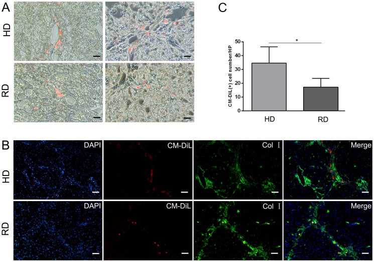
Figure 5. Distribution of transplanted cells in fibrotic liver.
(A) Transplanted cells with red fluorescence of 1,1′-dioctadecyl-3,3,3′,3′-tetra-methylindocarbocyanine dye (CM-DiL) were detected under confocal microscope (Scale bars: 50 µm). Positive cell were counted form five views from each sample with three samples in each group. *p<0.05. (B) Immunofluorescence staining of collagen type I (Col I) revealed that CM-DiL positive cells were located around portal vein and fibrous septa (Scale bars: 50 µm).

