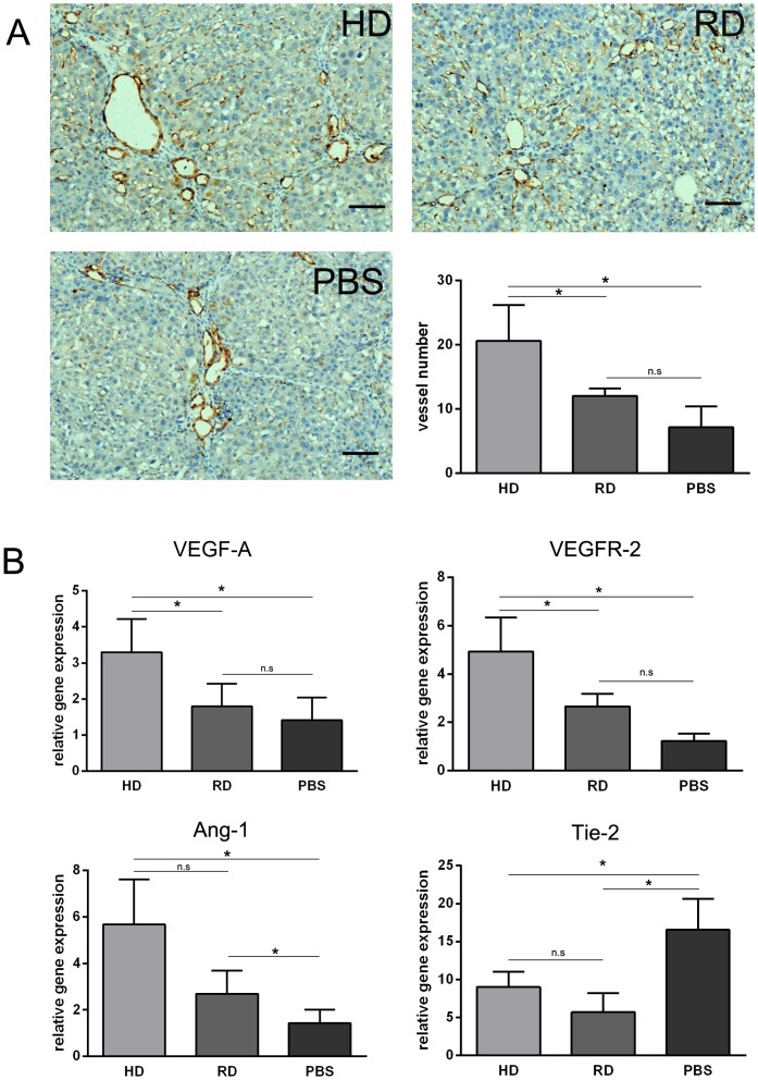
Figure 7. Increased sinusoidal blood vessel density by transplanted cells.
(A) Representative views of immunohistological staining of CD31 in HD group, RD group and PBS group (Scale bars: 150 µm). Number of CD31 positive vessels counted from immunohistological staining. Five views in each sample with three samples in each group were analyzed. (B) Expression of Ang-1, Tie-2, VEGF-A and VEGFR-2 analyzed by qRT-PCR. Each sample was repeated three times with three samples from each group. *p<0.05.

