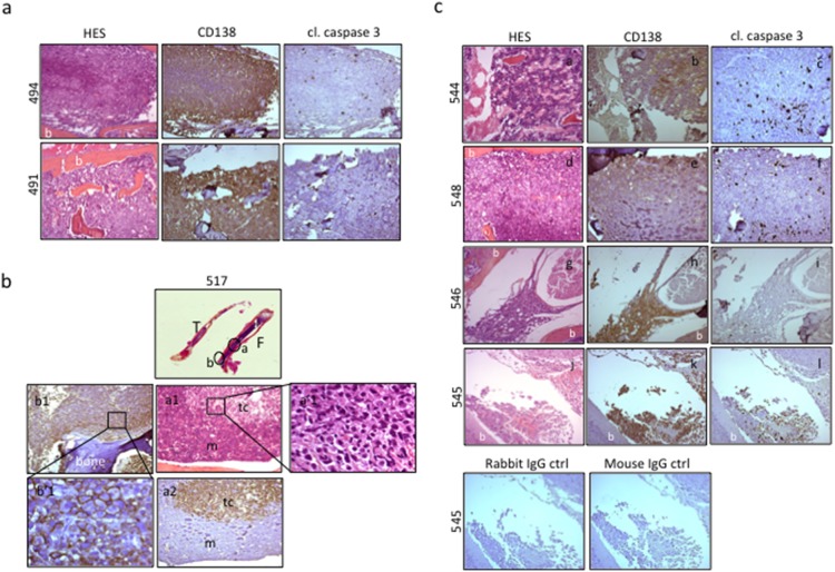
Figure 6. DZNep impairs the growth of MM cells in their niche.
(a) Bones from control mice were processed and analyzed by HES and by IHC for CD138 and cleaved (cl.) caspase 3 staining. Images (x100 magnification) obtained for the femur of mouse #494 and the rachis of mouse #491, both vehicle-treated. CD138-positive cells that invaded bone marrow, caused bone (b) disorganization within the femur (#494) and the intervertebral discs (#491). Concomitantly, few cl. caspase 3 staining was noticed. (b) The femur (F) and tibia (T) from vehicle-treated mouse #517 were processed, scanned (x30 magnification), HE stained and analyzed for CD138 labeling by IHC (x100 magnification, a1, a2, b1; x630 magnification, a’1, b’1). Foci (a, b, c, circled regions) of typical CD138-membrane stained (b’1) MM cells tumors cells (tc) are visible within disorganized mouse bone marrow (m). MM cells mainly concentrated in trabecular areas (b, b1, b’1) but also in delineated medullae foci (a, a1, a’1, a2). (c) Examples of histological analyses (HES, CD138 and cl. caspase 3 staining) from rachis (mice #544 and 546), femur (mouse #548) and skull (mouse #545) samples removed from DZNep-treated series. CD138-positive bona fide MM cells invaded bone (b) tissues causing destruction and disorganization (puddles or ghosts associated with elevated caspase 3 activity). This high caspase 3 activity underlined DZNep therapeutic efficacy. Images of negative isotype rabbit or mouse IgG controls done on skull sections from mouse 454 are shown.

