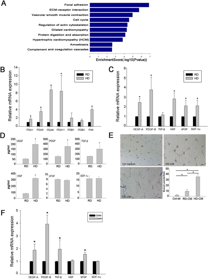
Figure 6. Up-regulation of cell adhesion molecules and pro-angiogenic growth factors in high density cultured cells.
A, Pathway analysis of microarray data. Genes involved in focal adhesion and ECM-receptor interactions were highly expressed in high density cultured cells compared with those in regular density cultured cells. B, Expression of integrin family genes validated by qRT-PCR analysis. Data are presented as the fold increase of gene expression in high density (HD) cultured cells compared with that in regular density (RD) cultured cells (n = 3). *p<0.05. C, Expression of growth factors validated by qRT-PCR analysis. Data are presented as the fold increase of gene expression in high density cultured cells compared with that in regular density cultured cells. (n = 3). *p<0.05. D, ELISA analysis of VEGF, PDGF, TGF-β, HGF, bFGF and SDF-1α in the supernatants of cultured cells. (n = 3). *p<0.05. E, Tube formation of HUVECs on Matrigel induced by conditioned medium from regular (CM-RD) or high density (CM-HD) cultures. Cells incubated in DMEM with 10% FBS served as a control medium (Ctrl-M). The number of branch points per field was counted after 12 hours of network formation (n = 9). *p<0.05. F, Growth factor expression of CD45+ and CD45− cells sorted from high density culture validated by qRT-PCR analysis. (n = 3). *p<0.05.

