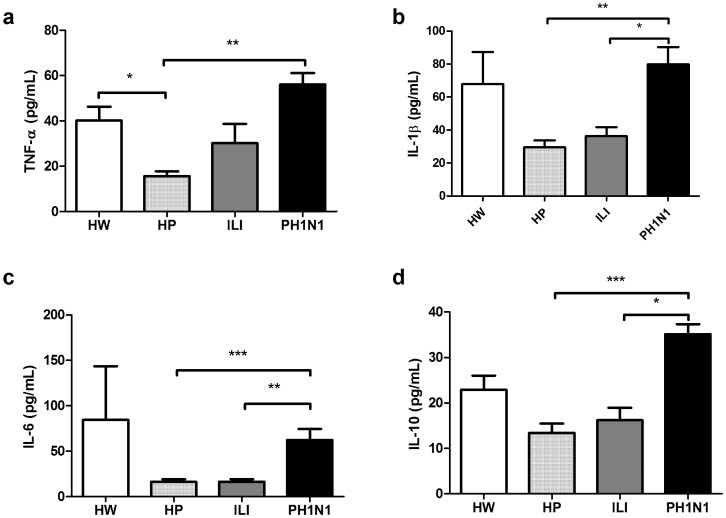
Figure 2. Serum cytokine concentrations in HW, HP, ILI and PH1N1 women.
Pro-inflammatory TNF-α (a), IL-1β (b) and IL-6 (c) and anti-inflammatory IL-10 (d) cytokines were quantified using a CBA system with flow cytometry. The Kruskal-Wallis test with Dunn’s multiple comparison post-test was performed using the GraphPad Software. The significance values were *p<0.05, **p<0.01, ***p<0.001.

