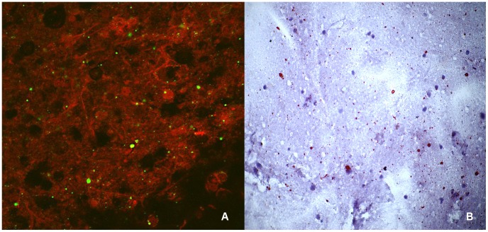
Figure 2. Touch impression of a rabies-positive domestic dog brain tested with the direct fluorescent antibody test (A) and direct rapid immunohistochemical test (B).
(A) Apple-green immunofluorescent viral inclusions observed on the red neuronal tissue in the brain processed by DFA. Magnification, ×400. (B) Magenta viral inclusions are visible on the blue neuronal background of the brain processed by dRIT. Magnification, ×200.

