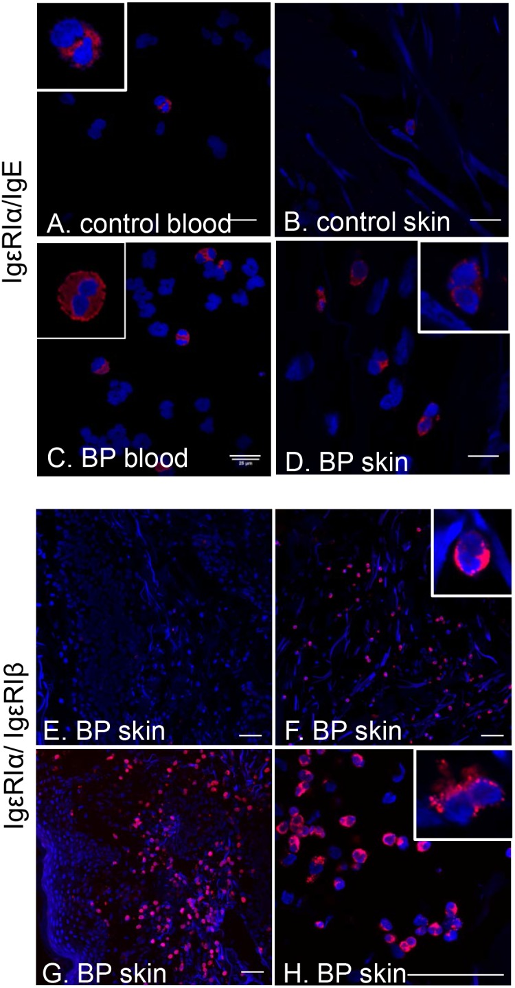
Figure 5. Surface expression of FcεRI on BP eosinophils.

Interaction of FcεRIα with IgE or FcεRIβ was evaluated using the proximity ligation assay on non-permeabilized preparations of circulating granulocytes or skin cryosections from BP patients or controls. Eosinophils were identified by their unique nuclear morphology (bi-lobed nucleus stained with DAPI) using high resolution confocal microscopy. Interaction of FcεRIα/IgE on peripheral blood and tissue eosinophils from BP patient (A, B) or controls (C, D). Insets are enlarged to show nuclear morphology. Scale bar = 25 uM. Interaction of FcεRIα/FcεRIβ on eosinophils in lesional biopsies (E–H). Panel H is an image of the same BP sample depicted in panel G, captured at higher magnification for resolution of nuclear morphology. Scale bar = 50 uM.
