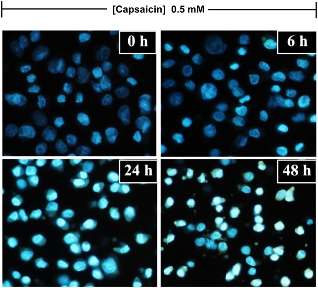
Figure 3. The effect of capsaicin on nuclear morphological alterations in HepG2 cells.
HepG2 cells were incubated with capsaicin at concentrations of 0.05 mM for 0, 6, 24 and 48 h. DAPI dye was used to indicate the apoptotic morphological features, such as chromatin condensation, nuclear fragmentation, and membrane blebbing, and then visualized by fluorescence microscopy at 40x magnifications. The control was defined as cells treated with a medium or vehicles without capsaicin (Scale bar: 25 µm).

