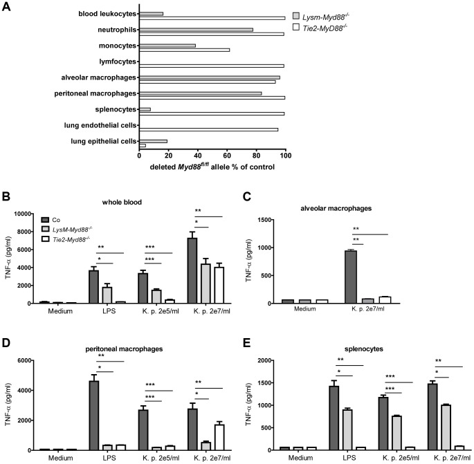
Figure 1. Genetic and functional characterization of primary cells from LysM-Myd88−/− and Tie2-Myd88−/− mice.
The residual amount of the MyD88fl/fl allel in blood and primary cells LysM-Myd88−/− and Tek-Myd88−/− mice was quantified via qRT-PCR relative to the unaffected Socs-3 gene. The amount of remaining “floxed” MyD88 region in LysM-MyD88−/− and Tek-MyD88−/− mice was calculated using the 2-deltaCt (ΔΔCt) method using the amount of genomic DNA from Myd88fl/fl mice for the no-deletion control. The deletion efficiency was calculated as (1 - residual Myd88fl) ×100% (A). Whole blood (B), alveolar and peritoneal macrophages (C,D) and splenocytes (E) derived from control, LysM-Myd88−/− and Tie2-Myd88−/− mice (n = 3 per group) were in vitro stimulated with LPS derived from Klebsiella pneumoniae (1 µg/ml) or heat killed K. pneumoniae in two concentrations (2×10⁵ CFU/ml or 2×10⁷/ml) for 20 hours. Data are expressed as mean (SE). * p<0.05, ** p<0.01, *** p<0.001.

