
Figure 4. SAM-II Superfamily.
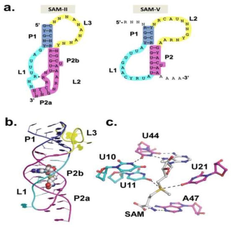
a) Consensus secondary structures of SAM-II and SAM-V riboswitch families[8]. The loops tolerate variability and long insertions. b) metX SAM-II crystal structure from Sargasso Sea metagenome[21]. Color scheme matches that in a. c.) SAM binding site in SAM-II. The Hoogsteen edge of SAM pairs with the Watson-Crick edge of U44 as part of a base triple. The SAM adenine stacks between G22 and A45. The sulfonium experiences favorable electrostatic interactions with U11 and U21 and the methionine tail hydrogen bonds with A47.