
Figure 5. SAM-III family.
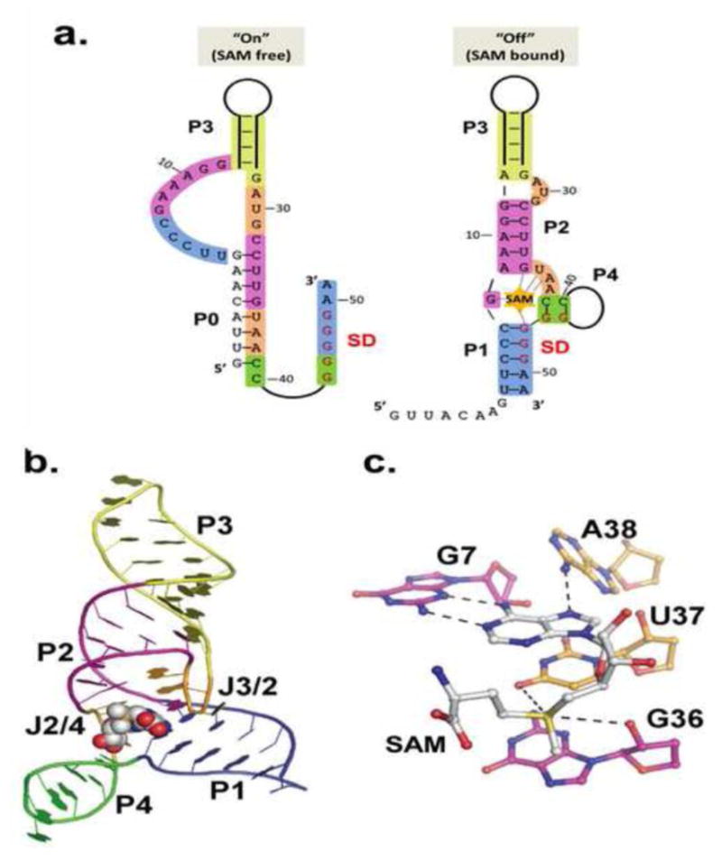
a.) Secondary structures of the on (SAM-free) and off (SAM-bound) states of the E. faecalis SAM-III riboswitch. Of the four secondary structure elements, only P3 is constant in both the on and off states. Bases involved in off state secondary structure elements are colored in both on and off state to emphasize changes. Coloring and numbering corresponds to off state crystal structure in B. b) Crystal structure of the SAM-bound E. faecalis SAM-III [23]. Coloring matches that in A. c.) SAM binding site in SAM-III. SAM stacks in the “binding pocket” between U37 (gold) and G48 (not shown). The adenosine pairs with G7 (purple) in the back of the pocket. Carbonyl oxygen atoms on U37 and G36 (in the SD) interact with the sulfonium.