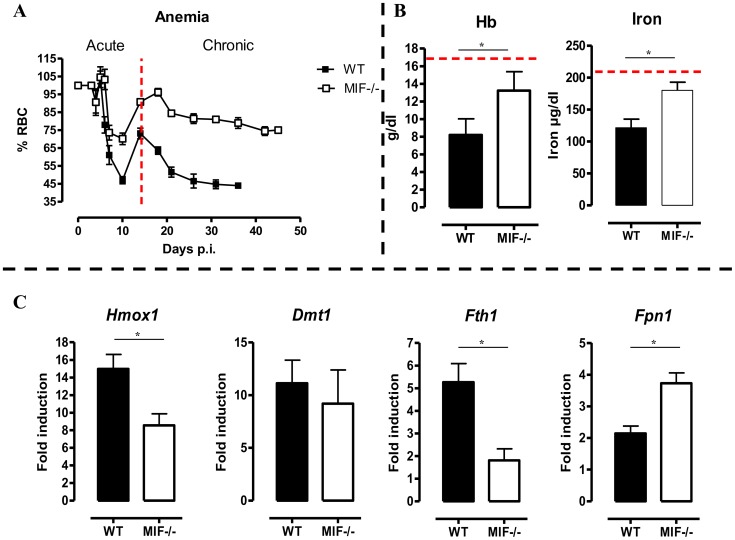
Figure 6. MIF deficiency correlates with reduced anemia, restored hemoglobin/serum iron levels and restored iron homeostasis during T. brucei infection.
(A) Anemia development during infection in C57Bl/6 (WT, black box; Mif −/−, white box) mice. Results are representative of 2–5 independent experiments and expressed as mean of 3–5 individual mice ± SEM. (B) At day 18 p.i., (left panel) hemoglobin levels and (right panel) serum iron levels in Mif −/− (open bars) and WT (black bars) mice. (C) Expression levels of the iron-homeostasis associated genes Hmox1 (iron import), Dmt1 (iron transport), Fth1 (iron storage) and Fpn1 (iron export) were quantified by RT-QPCR in total livers from Mif −/− (white bars) and WT (black bars) mice at day 18 p.i. Gene-expression levels are normalised using s12 and expressed relatively to expression levels of non-infected mice. Results are representative of 2 independent experiments and presented as mean of 3 individual mice ± SEM (*: p-values ≤0.05).

