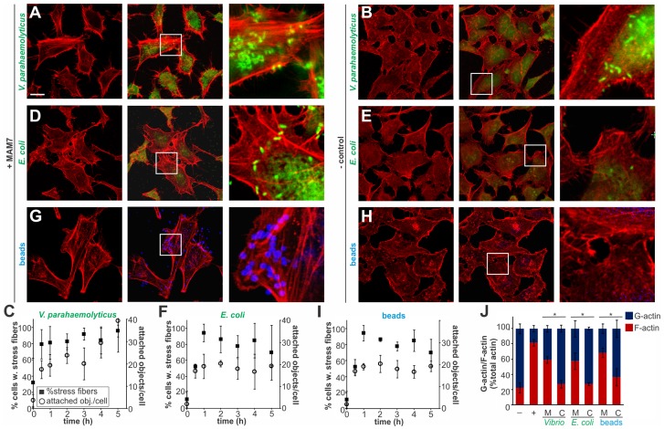
Figure 1. Clustering of MAM7 adhesin causes sustained actin rearrangements in host cells.
Attachment of V. parahaemolyticus CAB4 or E. coli BL21-MAM7 (A, D, bacteria expressing MAM7 in green) or of polymer beads coupled to GST-MAM7 (G, beads in blue) to Hela cells caused sustained stress fiber formation (F-actin stained with rhodamine phalloidin, red). Attached objects (bacteria or beads) per cell and % cells with stress fibers were determined from images taken at indicated timepoints (C, F, I). Data shown are means ± standard deviation from twelve images (4 frames from n = 3, representing at least 100 cells/experimental condition). V. parahaemolyticus CAB4ΔMAM7 or E. coli BL21-MAM7ΔN1–44 (MAM retained in the cytoplasm), (B, E, bacteria in green) or of polymer beads coupled to GST only (H, beads in blue) to Hela cells did not cause changes in the actin phenotype. Images shown are of 1 hour time points and are representative of a set of three experiments. Bar, 10 µm. G-actin (blue) and F-actin (red) content of cells treated with MAM (M) or controls (C) was quantified at 1 hour post treatment (J) and compared to serum-starved, untreated cells (−) and cells treated with F-actin enhancing solution (+). Results are means ± s.e.m. (n = 2) and (*) indicates statistical significance (p<0.05 in a student's two-tailed unpaired t-test).

