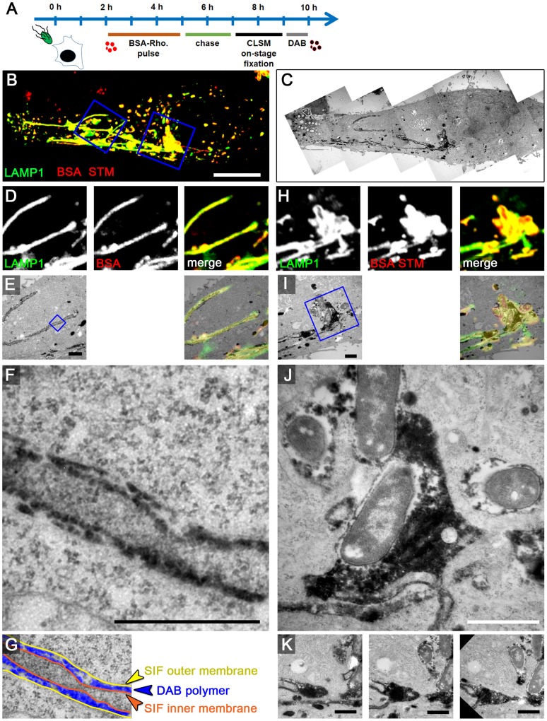
Figure 7. The outer lumen of double membrane SIF is in interchange with endocytosed material.
A) Scheme of the experiment. HeLa expressing LAMP1-GFP (green) were seeded on a petri dish with a gridded coverslip. Cells were infected with Salmonella WT expressing mCherry (STM, red). BSA-Rhodamine was added as fluid tracer to the medium 2–5 h p.i. After live cell imaging of selected cells at 8 h p.i. by CLSM (B, MIP), cells were immediately fixed on stage. DAB photo-conversion by Rhodamine was performed and cells were prepared for TEM. Several images of the same section were stitched for an overview (C). Details for LAMP1-positive, fluid tracer-labeled SIF (D–G) and SCV with attached SIF (H–K) are shown by correlative live cell CLSM (D, H, single Z plane) and TEM (E–G, I–K) micrographs. F) shows a higher magnification of double membrane SIF, and the pseudocolored micrograph (G) indicates the organization of inner and outer SIF membrane and the DAB polymer deposition in intermembrane lumen. J) shows DAP polymer deposition in direct contact to Salmonella within the SCV. Successive sections with several SIF extending from the SCV are shown in K). A cell representative of three biological replicates is shown (1–3 technical replicates with each 2–4 cells). Scale bars: 10 µm (B, C), 1 µm (D, E, H, I, K), 500 nm (F, J).

