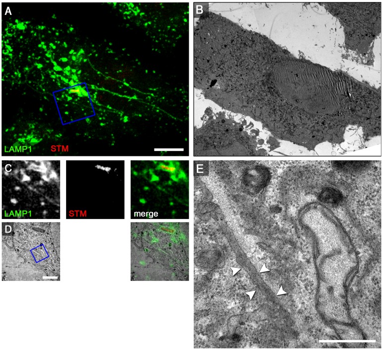
Figure 8. The SPI2-T3SS effector SseF is required for induction of double membrane SIF.
For CLEM analysis, HeLa cells stably transfected with LAMP1-GFP (green) were seeded on a Petri dish with a gridded coverslip and infected with the Salmonella sseF-deficient strain expressing mCherry (STM, red). After live cell imaging at 8 h p.i. by CLSM (A, MIP) cells were fixed immediately on stage. sseF-infected HeLa cells exhibit thin LAMP1-positive tubules. B) Low magnification TEM micrograph of the same cell. Details for LAMP1-positive thin SIF and Salmonella within SCV are shown by correlative live cell CLSM (C, F single Z plane) and TEM (D, G) micrograph. E, H) Higher magnifications of a SIF. The single membrane tubule is indicated by arrowheads. A cell representative of two biological replicates is shown (1–2 technical replicates with each 2–4 cells). Scale bars: 10 µm (A, B), 2 µm (C, D, F, G), 500 nm (E, H).

