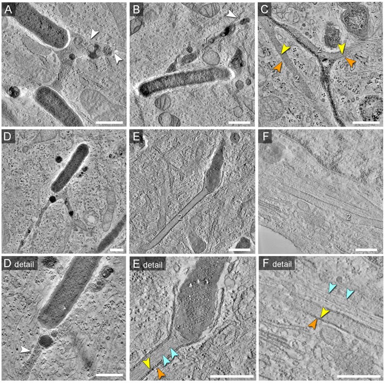
Figure 9. Morphology of SCV and SIT analyzed by EM tomography.
HeLa cells were infected with Salmonella WT as before, fixed and processed for EM tomography of partial cell volumes. A) to F) shows representative images corresponding to the tilt series shown in Movies S2, S3, S4, S5, S6, S7, S8, S9, S10. Note the presence of double membrane SIT indicated by orange and yellow arrowheads. SIT that are delimited by single membranes and that contain multi-lamellar vesicles and dense granules typical for late endosomes and lysosomes are referred to as type 1 SIT (A, B, D), with white arrowheads indicating SIT membranes. SIT that appear delimited by double membranes and lack multi-vesicular membranes are referred to as type 2 SIT (E, F). C) shows a ‘hybrid’ SIT resulting from partial fusion of two intertwined type 1 and type 2 SIT. E) shows a type 2 SIT with an inner space filled by a bundle of actin-like filaments, while SIT in F) are associated with F-actin filaments adjacent to the SIT (light blue arrowheads). Scale bars: 500 nm.

