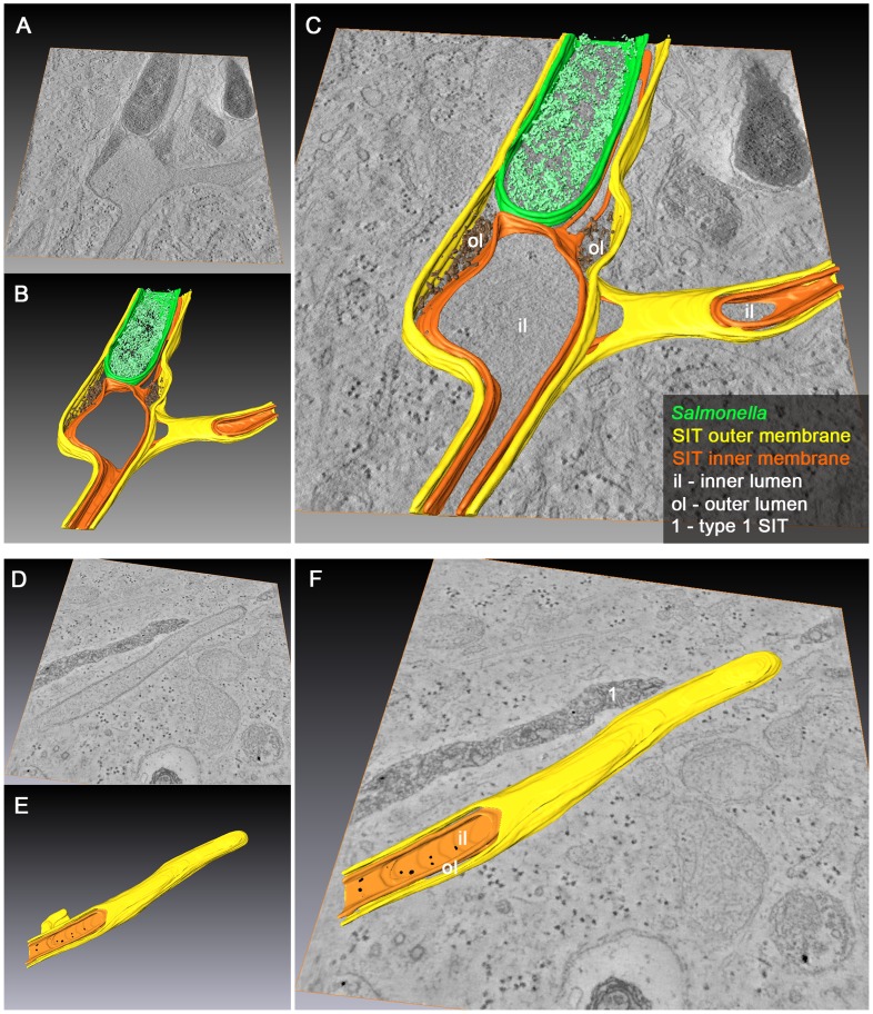
Figure 10. 3D organization of SCV and SIT in Salmonella-infected cells.
HeLa cells were infected with Salmonella WT for 10 h and processed for EM tomography of partial cell volumes followed by 3D-surface rendering. Representative images of single and double tilt series are shown in A) and D). A–C) Example of an SCV with extending and branching type 2 SIT showing dense luminal content between the two adjacent membranes at the base of SIT. D–F) Example of type 2 SIT with a closed tip and two membranes that wrap up portion of ribosome-containing cytosol. Note the presence of an adjacent type 1 SIT (without 3D-surface rendering) with a dense luminal content and delimited by a single membrane (E). The corresponding tilt series are shown in Movie S11 and Movie S13, respectively. B) and E) show 3D-surface rendering of the SIT and SCV membrane organization representing Movie S12 and Movie S14. Inner and outer SIT membranes are indicated in orange and yellow, respectively. The resulting inner and outer lumen of the double membrane tubular structure are labeled with iL and oL, respectively. Merged TEM images and rendered models are shown in C) and F).

