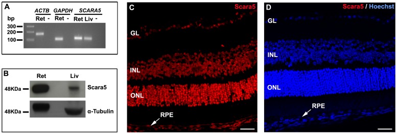
Figure 1. Scara5 was expressed in mouse retinal cells.
A: The expression of SCARA5 mRNA in the retina was evaluated by q-RT-PCR. Agarose gel electrophoresis of q-RT-PCR products confirmed that SCARA5 single amplicon with 108 bp was generated. ACTB and GAPDH were used as housekeeping genes. B: Western blotting analysis revealed a specific band with a molecular weight of 48 KDa, confirming the presence of Scara5 receptors in the retina. α-tubulin was used as a loading control. C: Retinal immunolabeling with a specific antibody in a histological section, along the eye axis through the optic disc and cornea, showed that Scara5 was expressed throughout the retina, mainly at the level of ganglion cell layer, inner nuclear layer, outer nuclear layer and RPE. D: Immunohistochemical negative control, where the primary antibody was omitted. Ret, retina; Liv, liver; -, no-template control; GL, ganglion cell layer; INL, inner nuclear layer; ONL, outer nuclear layer; RPE, retinal pigment epithelium. Scale bars: 28 µm (A); 28 µm (B).

