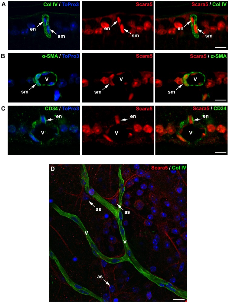
Figure 6. Retinal vasculature expressed Scara5 receptors.
A: Cells surrounded by the blood basement membrane marked with collagen IV showed intense Scara5 signal. B and C: Dual immunolabeling with Scara5 and with α-SMA and CD34, respectively, confirmed that vascular smooth muscle cells and endothelial cells expressed Scara5 receptors. D: Whole mount retinas immunohistochemically marked with collagen IV and Scara5 showed that perivascular astrocyte-like cells intensively expressed Scara5 in their vascular end-feet. Nuclei were counterstained with ToPro3. en, endothelial cell; sm, smooth muscle cell; v, blood vessel; as: astrocyte-like cell. Scale bars: 10 µm (A); 9 µm (B); 9 µm (C); 12 µm (D).

