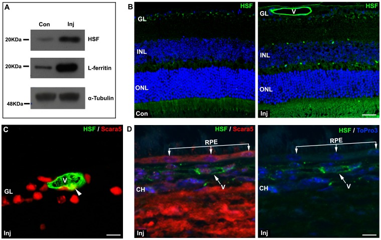
Figure 7. Intravenously injected HSF crossed the BRB.
A: Six hours after the intravenous injection of HSF, western blotting analysis revealed that HSF was present in the retina. As expected, a marked increase of L-ferritin content was also confirmed. B: The immunolabeling with a specific anti-HSF antibody showed that HSF crossed the inner BRB and accumulated in mouse retina. HSF was internally lining the retinal blood vessels. C and D: The double staining with anti-HSF and with anti-Scara5 antibodies showed that L-ferritin co-localized with endothelial cytoplasmic Scara5 (arrowhead), but no content of HSF was observed in RPE cells, suggesting a differential function of the inner and outer component of BRB. Nuclei were counterstained with ToPro3. Con, non-injected control; Inj, injected; V, blood vessel; GL, ganglion cell layer; INL, inner nuclear layer; ONL, outer nuclear layer; RPE, retinal pigment epithelium; CH, choroid. Scale bars: 24 µm (B); 10 µm (C); 8 µm (D).

