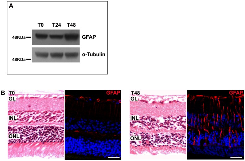
Figure 9. Murine model of retinopathy with photoreceptor degeneration.
A: Forty-eight hours after sodium iodate injection, retinas analyzed by western blotting showed an increased expression of GFAP. α-tubulin was used as a loading control. B: Paraffin-embedded retinal sections stained with hematoxylin-eosin or immunolabeled with a specific anti-GFAP antibody revealed photoreceptor alterations and gliosis, indicating that retinopathy was well established. Nuclei were counterstained with ToPro3. GL, ganglion cell layer; INL, inner nuclear layer; ONL, outer nuclear layer. Scale bar: 35 µm.

