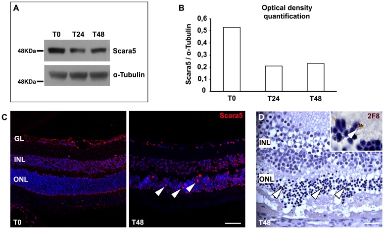
Figure 10. Scara5 expression decreased during retinopathy.
A: Retinas of a sodium iodate murine model were analyzed by western blotting. Scara5 expression reduced to about half during the treatment. B: Graph representing the optical density quantification of the Western blotting analysis for Scara5, after normalization with respect to α-tubulin. C: The immunolabeling of paraffin-embedded retinal sections with anti-Scara5 antibody confirmed that Scara5 expression decreased throughout the parenchyma. However, several positive Scara5 cells were found in the outer nuclear layer (arrowhead). D: During retinopathy, 2F8 positive cells were also observed in the outer nuclear layer (arrowhead), with a disposition compatible with that of Scara5 positive cells. 2F8 was revealed by DAB reaction and histological sections were counterstained with hematoxylin. GL, ganglion cell layer; INL, inner nuclear layer; ONL, outer nuclear layer. Scale bars: 45 µm (C); 20 µm (D).

