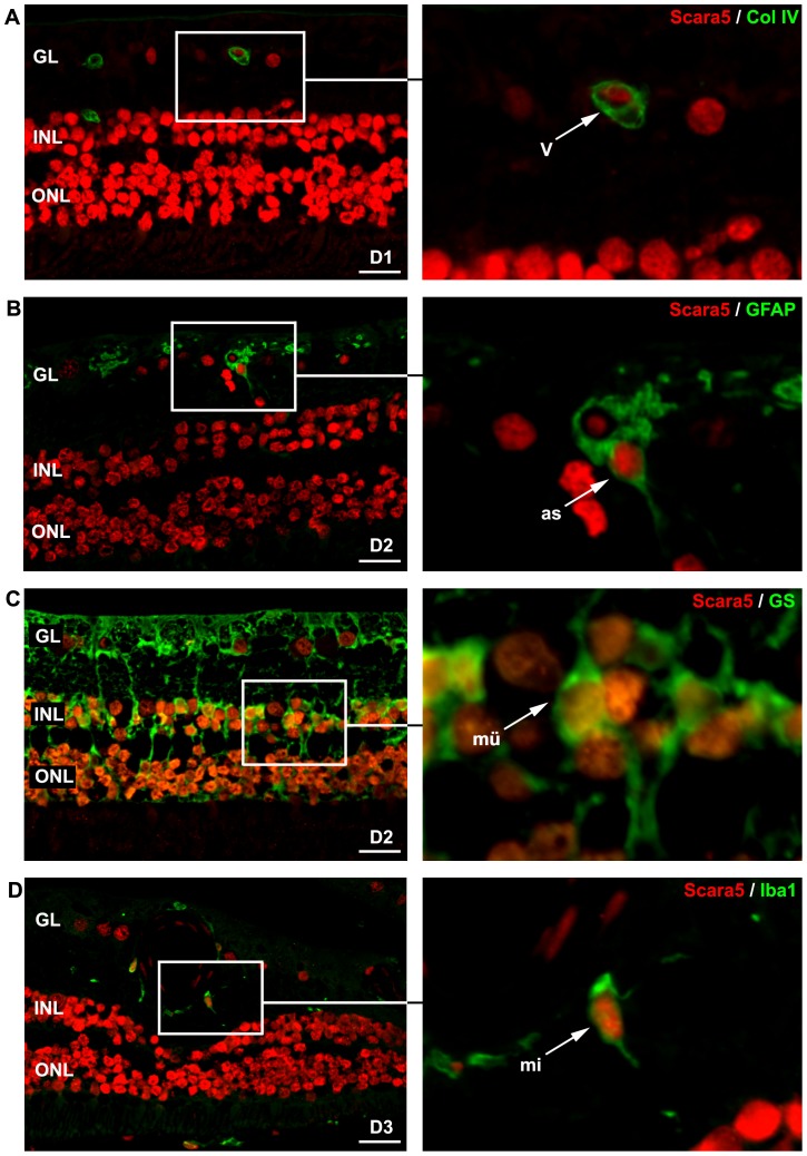
Figure 11. Scara5 was expressed in human retinal cells.
Laser confocal analysis of double-stained paraffin-embedded human retinal sections with anti-Scara5 and anti-collagen IV, anti-GFAP, anti-GS and anti-Iba1 antibodies revealed that Scara5 was present throughout the retina, including endothelial cells, astroctyes, Müller cells and microglial cells, following the distribution pattern observed in mouse retinas. D1, D2 and D3, 42, 78 and 86-years-old healthy human donors, respectively; GL, ganglion cell layer; INL, inner nuclear layer; ONL, outer nuclear layer.: 22 µm (A); 21 µm (B); 24 µm (C); 28 µm (D).

