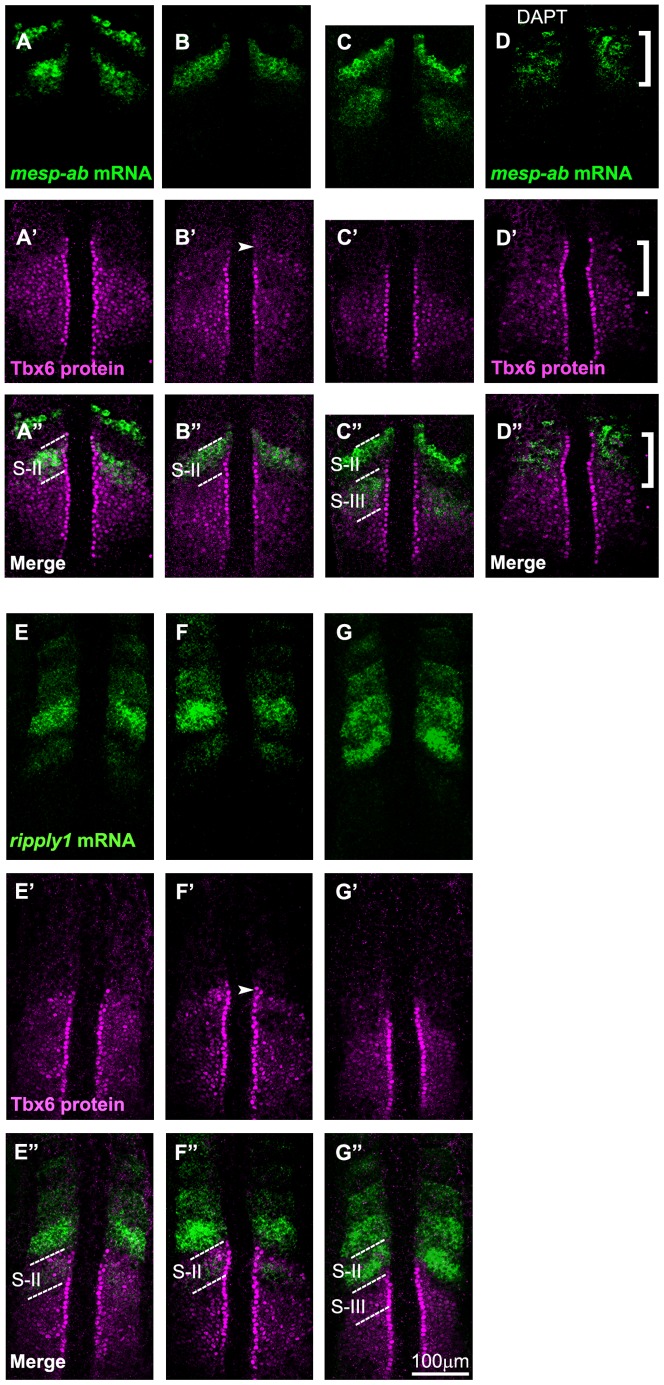
Figure 3. Comparative analysis of the anterior border of the Tbx6 domain with expression of mesp and ripply.
Spatial pattern of the Tbx6 protein (magenta) is compared with those (green) of mesp-ab mRNA (A-D) and ripply1 mRNA (E-G) at 3 different phases of segmentation cycle (the phases in embryos shown in A, B, or C are identical to those shown in E, F, or G, respectively) at around the 8 somite stage. Tbx6 pattern was also compared with mesp-ab mRNA in embryos treated with DAPT (D). Merged images are indicated (A″-G″). Out of a total of 59 embryos examined, around 46.5% of them showed A and E phase, 27% showed B and F phase, 26.5% showed C and G phase type of expression. Note that the anterior limit of newly expressed, or most posterior, mesp-ab band coincided with the anterior border of the Tbx6 core domain (A-A″). Then, this expression coincided with the upper band of Tbx6 when elimination of the anterior Tbx6 domain started (B-B″). On the other hand, new ripply1 expression emerged within the anterior part of Tbx6 domain (E-E″) and the Tbx6 domain starts to vanish in area where ripply1 was expressed (F-F″). In (D), the defects were observed in all of the embryos treated with DAPT (n = 22). Patterns of Tbx6 proteins and mesp-ab mRNA were disturbed in anterior area indicated by a bracket. The dotted lines indicate S-II (A″, B″, E″, F″) and, S-II and S-III (C″, G″) regions.

