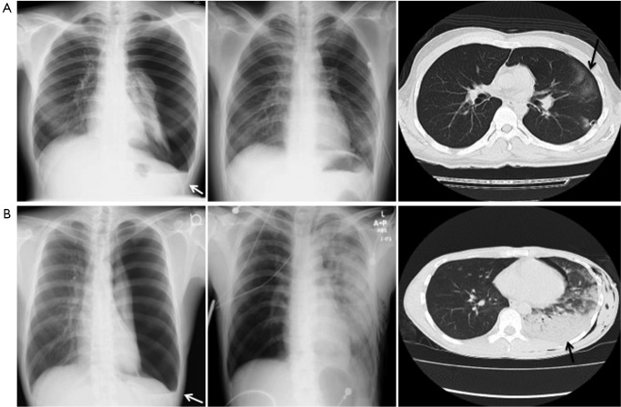
Figure 1.
Representative chest radiographs and CT scan of a pneumothorax that was associated with the development of asymptomatic RPE (A) and symptomatic RPE (B). Both figures show X-rays of pleural effusion (white arrow) in addition to pneumothorax (left column). Both of the X-rays taken after tube thoracostomy reveals a hazy ground-glass infiltrate in the left lower-lobe (middle column). Right column indicates CT images of RPE (black allow); RPE, reexpansion pulmonary edema.

