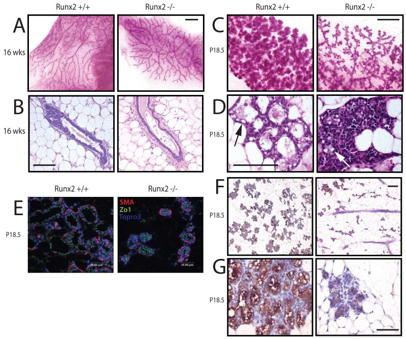
Figure 2.
Failed lobuloalveolar development in Runx2−/− mammary glands.
A) Whole mounts of transplanted Runx2+/+ and Runx2−/− mammary glands isolated from virgin Rag1−/− recipients at 16 weeks post-transplantation (Scale bar 1 mm).
B) H&E stained section through epithelial duct of glands shown in (A) (Scale bar 12.5 μm).
C-D) Same as in (A-B), but transplanted mammary glands collected at day 18.5 of pregnancy (P18.5) Arrows indicate lipid globules.
E) Sections of P18.5 Runx2+/+ and Runx2−/− mammary glands were analysed by immunofluorescence for polarity markers ZO1 (apical) and smooth muscle actin (SMA: basal).
F) Immunohistochemical analysis of β casein expression in Runx2+/+ and Runx2−/− mammary glands at P18.5 (Scale bar 25 μm). Higher magnification image shown in (G).

