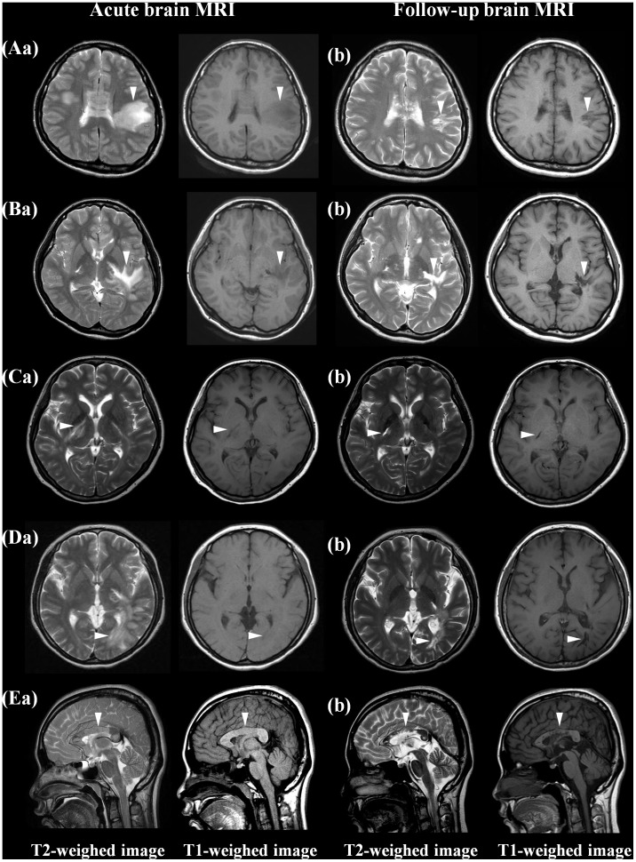
Figure 2. Chronic cystic changes of brain lesions on MRI.
At the time of an acute brain attack, brain MRI showed multiple T2-hyperintense lesions with subtle T1 hypointensity in the frontal white matter (Aa), corticospinal tract (Ba, Ca), occipital white matter (Da) and corpus callosum (Ea). On follow-up MRI, all T2 hyperintense lesions were markedly decreased in size but revealed focal T1-hypointensity with cystic changes (Ab, Bb, Cb, Db, Eb).

