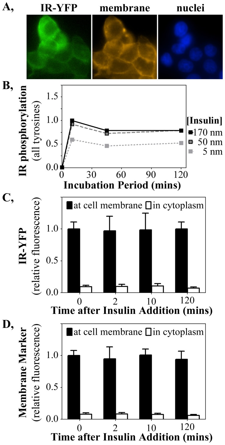
Figure 5. The IR remains membrane localized following chronic insulin stimulation of HTC-IR-YFP cells.
(A) YFP fused to the IR fluoresces green and co-localizes with the membrane-specific stain (orange) and not with nuclei (blue). (B) Total IR tyrosine phosphorylation in HTC-IR-YFP cells, detected by ELISA, mirrors that of the unfused similar to HTC-IR in response to insulin stimulation (see Fig. 1D). The fluorescence intensity of (C) YFP (IR) and (D) the membrane marker were quantified at the membrane and in the cytosol of HTC-IR-YFP cells.

