
Figure 3. Oncopeltus fasciatus hatched from eggs submitted to surface asepsis were Leptomonas wallacei-free.
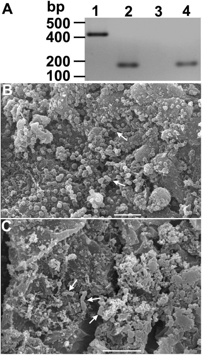
(A) Representative gel electrophoresis of PCR-amplified DNA samples extracted from eggs and whole insect guts. Lane 1- The DNA extracted from an axenic culture of L. wallacei was amplified with primers specific for parasite detection. Lane 2- Sample of pooled DNA extracted from eggs collected at the infected colony and submitted to surface asepsis was concomitantly amplified with primers specific for parasite or insect DNA detection. Lanes 3 and 4- Sample of pooled DNA, extracted from 3 pools of five insect guts of insects hatched from eggs submitted to asepsis, was PCR-amplified with primers specific for parasite (lane 3) or insect DNA (lane 4) detection, respectively. On the left, the positions of molecular size markers are shown in base pairs. The figure represents a negative image of the gel. (B and C) Scanning electron microscopy of O. fasciatus midgut and hindgut, respectively. The micrographs show the presence of bacteria (arrows) but absence of parasites. Bars = 10 µm.