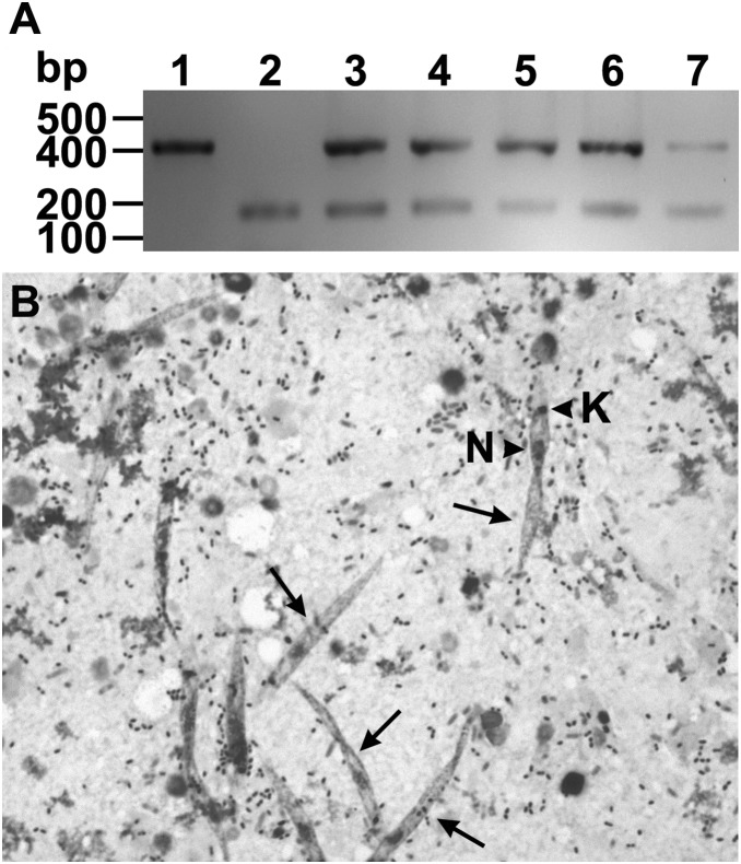
Figure 4. Detection of parasites after experimental transovum transmission of Leptomonas wallacei parasites by Oncopeltus fasciatus.
(A) Detection of parasite infection by PCR. Lane 1- The DNA extracted from an axenic culture of L. wallacei was amplified with primers specific for parasite detection. Lane 2- A pool of DNA samples extracted from gut of parasite-free insects was concomitantly amplified with primers specific for parasite and insect DNA detection. Lanes 3–7- Representative DNA samples extracted from guts of insects that fed on sunflower seeds contaminated with eggshels collected from infected colony were concomitantly amplified with primers specific for parasite and insect DNA detection. On the left, the positions of molecular size markers are shown in base pairs. The figure represents a negative image of the gel. (B) Detection of parasite infection by optical microscopy. The representative micrograph shows Giemsa-stained parasites (arrows) in the gut contents of newly infected insects. The arrowheads indicate the nucleus (N) and kinetoplast (K) of the parasite. Magnification = 400 x.

