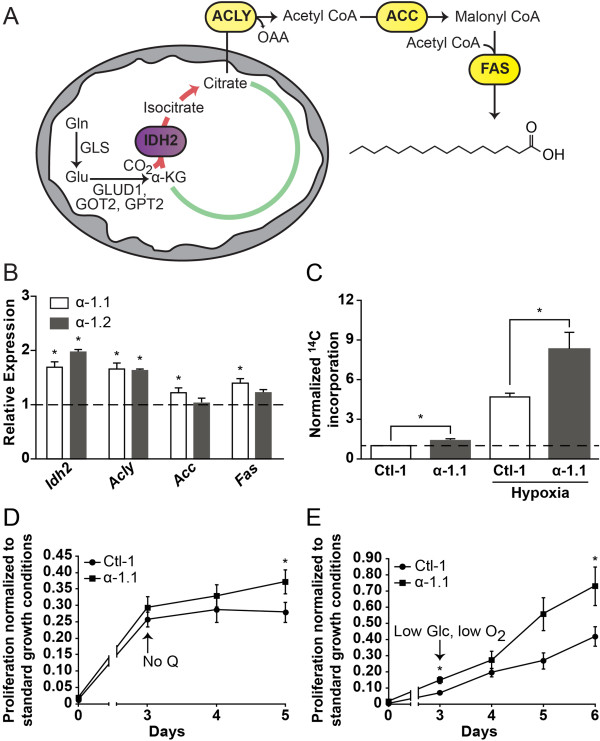
Figure 4.

PGC-1α promotes glutamine-mediated lipid biosynthesis and favors proliferation in limited nutrient conditions. (A) Schematic representation depicting lipogenesis from glutamine through reverse citric acid cycle (CAC) activity. Glutamine-derived α-ketoglutarate is converted to isocitrate by mitochondrial NADP+/NADPH-dependent isocitrate dehydrogenase (IDH2), producing citrate. Citrate is exported to the cytoplasm to generate acetyl CoA and oxaloacetate via ATP citrate lyase (ACLY). Acetyl CoA is converted to malonyl CoA by acetyl CoA carboxylase (ACC). Iterative additions of acetyl CoA to malonyl CoA by fatty acid synthase (FAS) yield fatty acid products (shown: palmitate). Gln, glutamine; Glu, glutamate; OAA, oxaloacetate; GLS1, kidney-type glutaminase; GLS2, liver-type glutaminase; GLUD1, glutamate dehydrogenase 1; GOT2, mitochondrial glutamic-oxaloacetic transaminase; GPT2, mitochondrial glutamic pyruvate transaminase. (B) Expression of lipogenic genes in ERBB2/Neu-induced breast cancer cells (NT2196) with increased expression of PGC-1α (α-1.1, α-1.2) normalized to that in control cells. Data are presented as means ± S.E.M., n = 3. *P <0.05, paired Student's t-test. (C) Incorporation of 14C into lipids from trace [U-14C]-glutamine in α-1.1 and Ctl-1 cells under normoxia or hypoxia. Counts were normalized for cell number and expressed relative to that of control cells in normoxia. Data are presented as means ± S.E.M., n = 6. *P <0.05, paired Student's t-test. (D) Relative proliferation of α-1.1 and Ctl-1 cells in the absence of glutamine compared to proliferation in standard glutamine conditions. Data are presented as fold values of cell count in standard glutamine conditions at 5 days ± S.E.M., n = 6. *P <0.05, paired Student's t-test. (E) Relative proliferation of α-1.1 and Ctl-1 cells in hypoxia under low glucose conditions compared to proliferation in normoxia and standard glucose conditions. Data are presented as fold values of cell count in normoxia and standard glucose conditions at 6 days ± S.E.M., n = 4. *P <0.05, paired Student's t-test.
