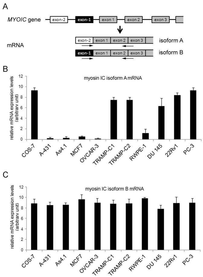
Figure 2. mRNA expression of myosin IC isoforms A and B in mammalian cell lines.
Detection of myosin IC isoforms A and B mRNA levels in various mammalian cell lines by qRT-PCR using isoform-specific primer. A) Schematic depicting the 5′ region of the MYOIC gene and the resulting isoform A and B mRNAs. The target sequence location of primer used for qRT-PCR to detect myosin IC isoforms are indicated by arrows. B) Quantitative real-time PCR analysis of mRNAs expression levels of myosin IC isoform A normalized to GAPDH. C) Quantitative real-time PCR analysis of mRNAs expression levels of myosin IC isoform B normalized to GAPDH. Myosin IC isoform A mRNA expression is high in COS-7 cells and the human and mouse prostate cancer cell lines PC-3 and TRAMP-C2. In contrast, myosin IC isoform B mRNA is expressed at comparable levels in all analyzed ell lines. Results are presented as means ± standard deviation; n = 3 independent experiments.

