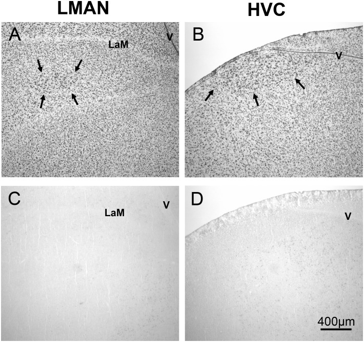
Figure 6. Relative absence of ZENK expressing cells in LMAN and HVC.
Panels A and B depict thionin stained sections, with arrows showing borders of LMAN and HVC from a male exposed to arrhythmic song. Panels C and D depict adjacent sections with representative samples of immunohistochemical labeling. LaM = lamina mesopallialis; V = ventricle.

