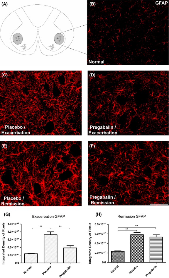
Figure 5.

(A) Diagram showing the region of lamina IX where the motoneurons are present. (B) Normal GFAP-1 labeling. (C) Increase of GFAP immunoreactivity in the placebo group, especially in the neuropil adjacent to motoneurons, during peak disease. (D) Amelioration of the astrogliosis following pregabalin treatment. (E and F) Anti-GFAP labeling at the remission phase. Note that there is no difference between placebo- and pregabalin-treated groups. (G) Quantification of the immunolabeling in the different experimental groups during peak disease. (H) Quantification of the immunolabeling in the different experimental groups during remission phase. *P < 0.05, **P < 0.01, ***P < 0.001. N = 6 for each experimental group. Scale bar 50 μm.
