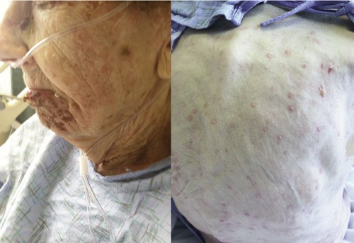
Figure 1.

Clinical photograph of a patient with disseminated cutaneous herpes zoster. On the left, cluster of vesicular lesions involving the mandibular branch of the trigeminal nerve. On the right, diffuse vesicular rash with erythematous base involving the back.
