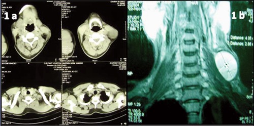
Figure 1.

a) Computed tomography neck and thorax showed a well defined cystic lesion in the posterior triangle in relation to middle one-third of sternocleidomastoid having thin wall and internal septations. b) Magnetic resonance imaging of soft tissue neck was suggestive of third branchial cleft cyst.
