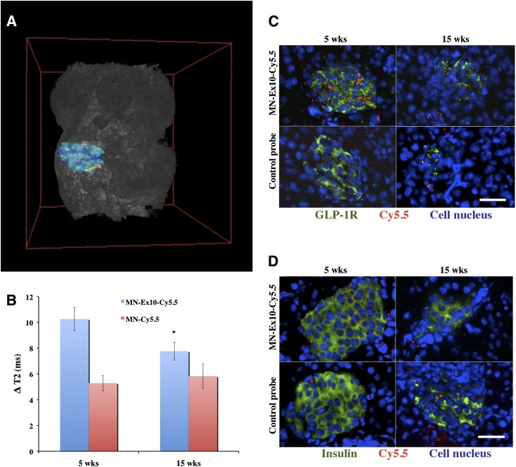
Figure 4.
T2-weighted MRI in NOD mice. A: Representative 3D T2*-weighted MRI of a 5-week-old NOD mouse shows pancreatic area used for analysis. B: The ΔT2 of the pancreata of 15-week-old mice injected with MN-Ex10-Cy5.5 was significantly decreased (*P < 0.05) compared with that of 5-week-old NOD mice. C: Anti–GLP-1R immunostaining of the pancreata from prediabetic and diabetic NOD mice injected with MN-Ex-Cy5.5 or control probes. Cy5.5 (red), GLP-1R (green), DAPI nuclear stain (blue); magnification bar = 20 μm. D: Anti-insulin immunostaining of the pancreata from prediabetic and diabetic NOD mice injected with MN-Ex-Cy5.5 or control probes. Cy5.5 (red), insulin (green), and DAPI nuclear stain (blue); magnification bar = 20 μm. wks, weeks.

