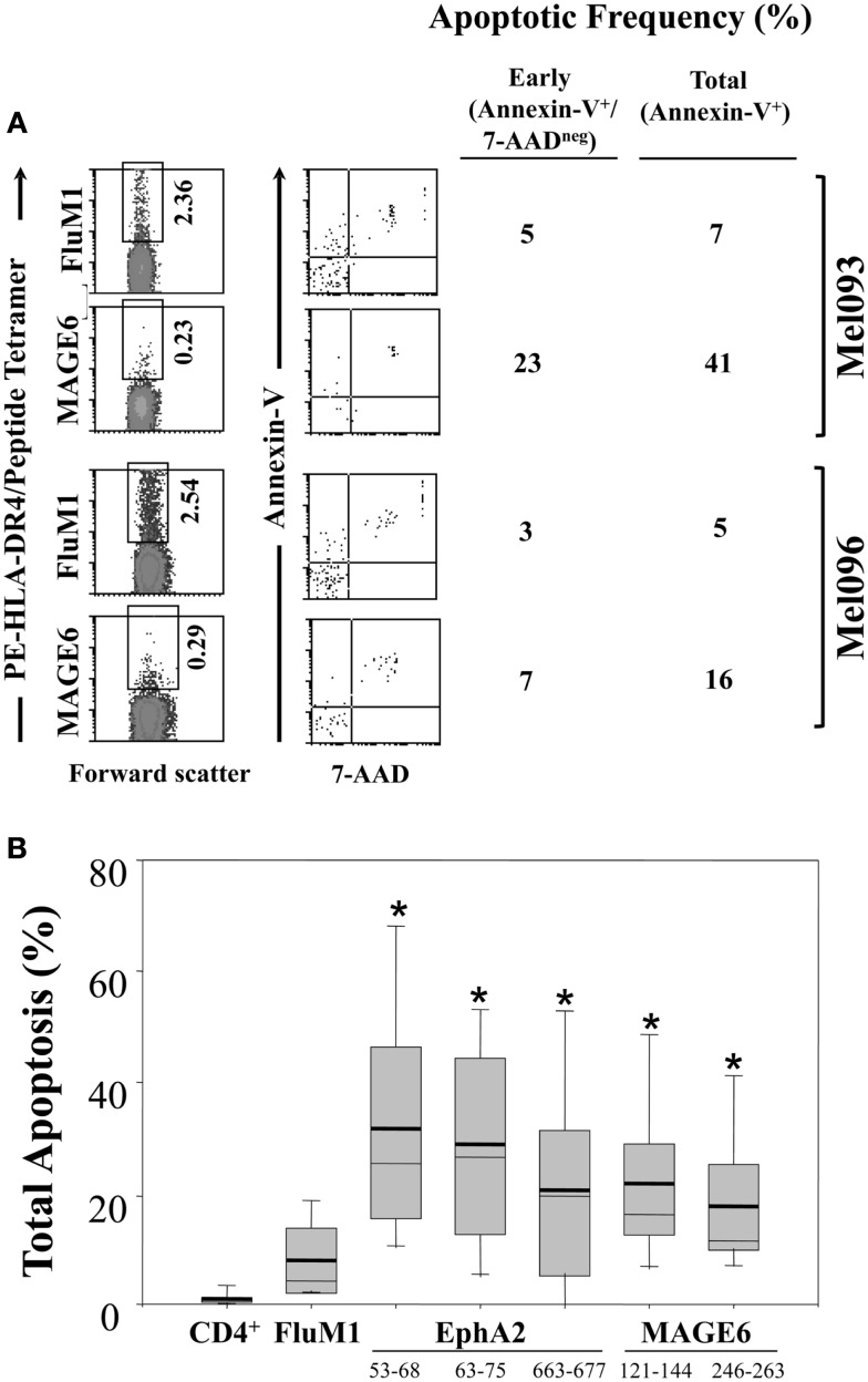
Figure 4.
Enhanced apoptosis of TAA (vs. FluM1)-specific CD4+ T cells in the peripheral blood of melanoma patients. Flow cytometric analysis of CD4+ HLA-DR4/peptide tetramer+ T cells was performed as described in Figure 1. (A) Peripheral blood antigen-specific (tetramer+) CD4+ T cells from patients Mel093 and Mel096 were apoptotic status based on staining for Annexin-V and 7-AAD as monitored by multi-parameter flow cytometry (as described in Section “Materials and Methods”). Frequencies of early (Annexin-V+/7-AADneg) vs. total (Annexin-V+ regardless of 7-AAD status) apoptotic events is tabulated. (B) Cumulative results for total CD4+ T cells and antigen-specific CD4+ T cells are depicted in Box plot format, with heavy horizontal lines representing the mean for each group, and whisker bars representing 5th and 95th percentiles (n = 13; i.e., Mel036-Mel104). *p < 0.05 vs. FluM1 (T-test).

