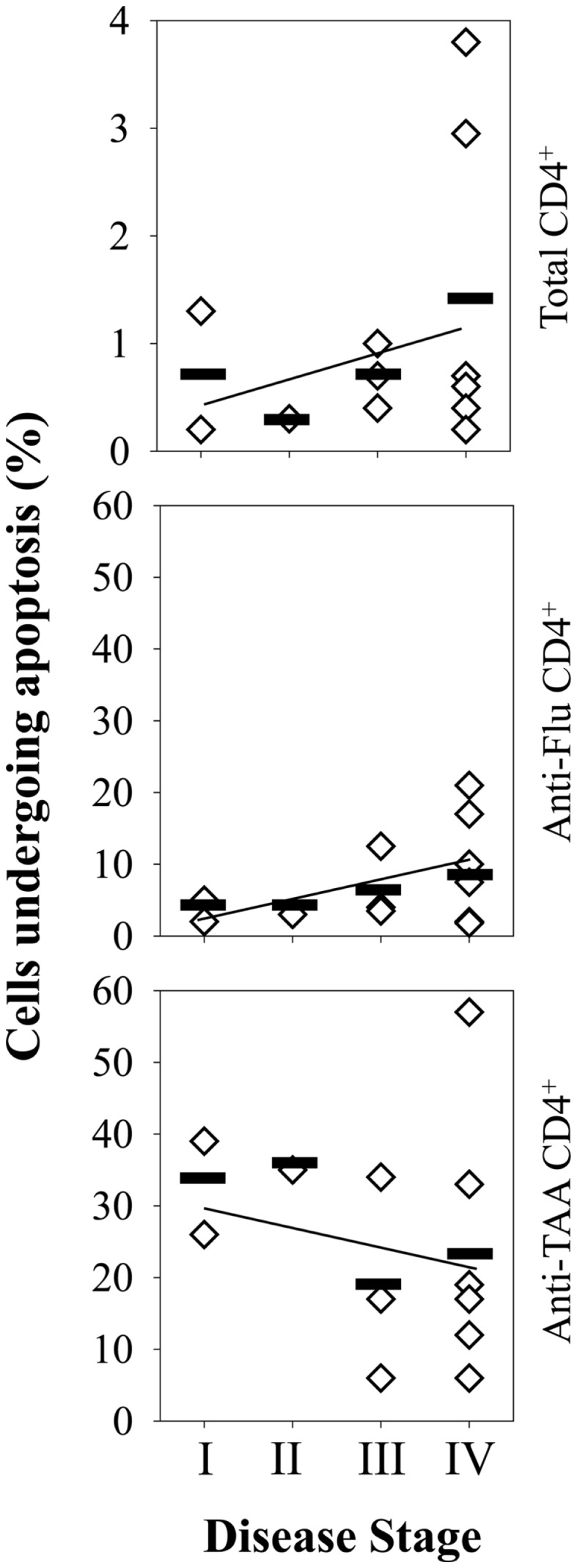
Figure 5.

TAA-specific CD4+ T cells in the peripheral blood of melanoma patients undergo enhanced rates of apoptosis (vs. Flu-M1-specific CD4+ T cells) at all stages of disease progression. Frequencies of total apoptotic events among total CD4+ T cells and antigen-specific CD4+ T cells in melanoma patient PBMC (n = 13) were determined using PE-labeled HLA-DR4/peptide (pooled TAA or FluM1) tetramers and flow cytometry as described in Figure 4. Patients were segregated based on clinical disease stage, with each diamond symbol representing data from a single patient. Linear-regression trend lines overlay each graph.
