Figure 5.
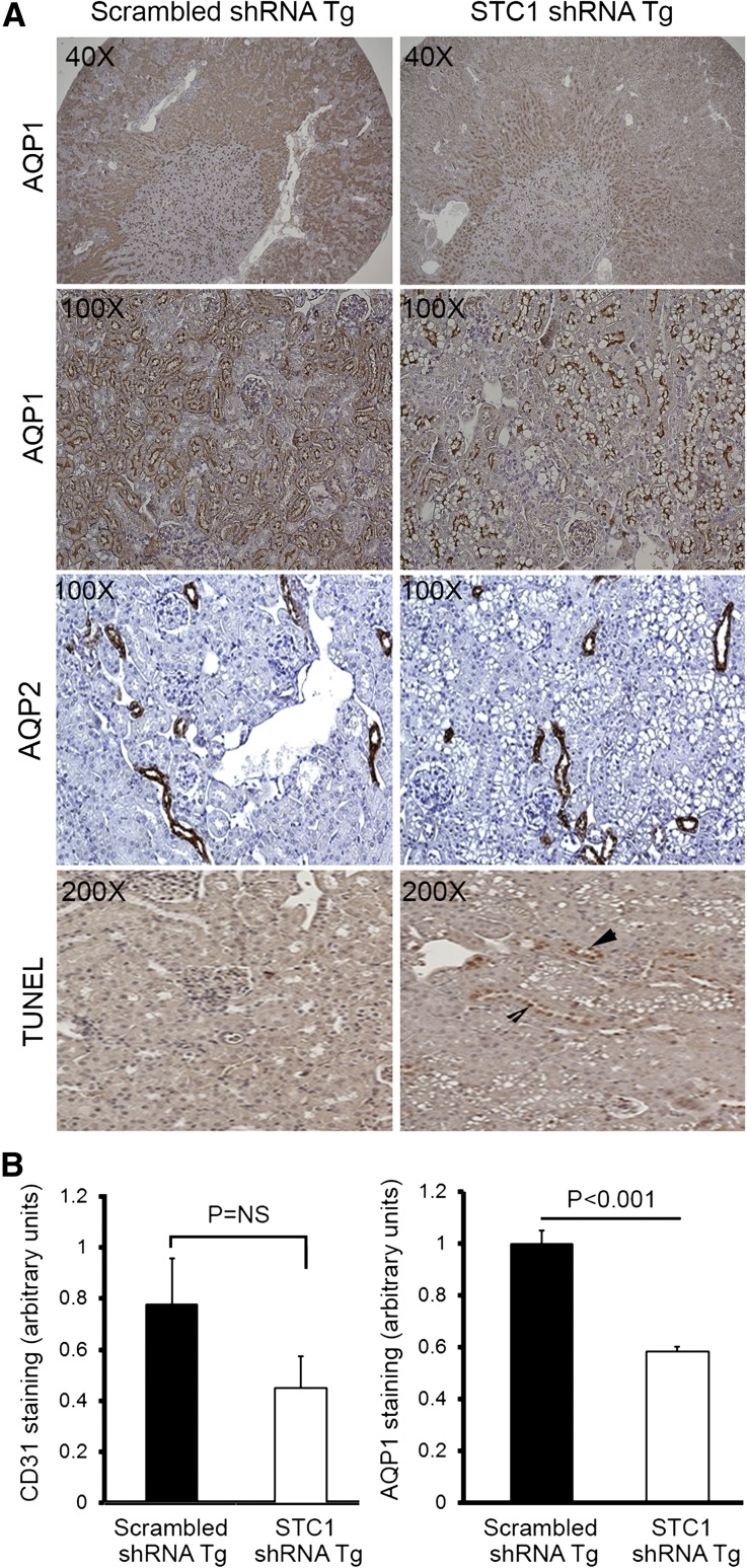
(A) STC1 knockdown induces vacuolization in S1 and S2 segments of the PT and promotes apoptosis. pTie2-Cre was delivered to the right kidneys of STC1 shRNA and scrambled shRNA Tg mice. Kidneys were harvested 4 days later and fixed in methacarn. Sections were stained with rabbit anti-AQP1, rabbit anti-AQP2, or TUNEL for apoptosis. Note that STC1 shRNA Tg kidneys display selective injury (vacuolization) in AQP1-positive tubules, in the outer two thirds of the cortex, with relative preservation of AQP1 staining in the inner third, consistent with tubular injury in the S1 and S2 segments of the PT. No vacuolization is observed in AQP2-expressing cells. Apoptotic cells (arrowheads) are more prevalent in STC1 shRNA Tg kidneys. (B) Apparent decrease in AQP1 and CD31 staining after STC1 knockdown does not correlate with decrease in protein expression (refer to C). Bar graphs show quantitation of staining for CD31 and AQP1 (using Image Tool software) 4 days after the delivery of Tie2-Cre to scrambled shRNA and STC1 shRNA Tg kidneys. (C) STC1 knockdown diminishes the expression of UCP2, but not AQP1 and CD31 proteins. Four days after the delivery of Tie2-Cre to scrambled shRNA and STC1 shRNA Tg kidneys, equal amounts of cortical homogenate in RIPA buffer were run on SDS-PAGE and blots were reacted with antibodies for CD31, UCP2, AQP1, and actin. Representative blots are shown. Bar graphs show the mean±SEM of band intensities for the above proteins normalized to actin; data represent six independent determinations.


