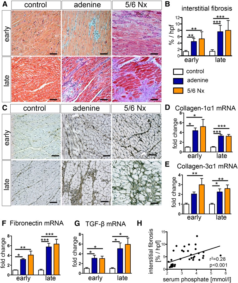
Figure 3.
Uremic cardiomyopathy is characterized by interstitial myocardial fibrosis with upregulation of TGF-β expression. (A and B) Trichrome staining of hearts (A) and quantification (B) demonstrates significantly increased interstitial myocardial fibrosis in both models of UC at early and late time points. (C and D) Staining (C) and quantitative real-time PCR (D) for collagen 1α1 revealed increased expression in both rat models of UC. (E and F) Other extracellular matrix proteins, such as collagen 3α1 (E) and fibronectin (F), also showed increased expression of mRNA and protein levels (immunostaining: Supplemental Figure 1). (G) Quantitative real-time PCR showed increase expression of TGF-β mRNA transcript in both rat models of UC at early and late time points. (H) The severity of interstitial myocardial fibrosis did correlate significantly with serum phosphate levels. *P<0.05; **P<0.01; ***P<0.001 (one-way ANOVA with post hoc Scheffe test). All scale bars=50 μm.

